
 1")
Description
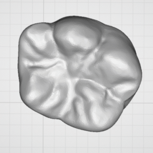
Mandibular Molar 3D Model
Watertight STL of a mandibular molar with a continuous root canal and simulated open apices. Built for training MTA apical plug/apical barrier techniques, conservative access cavity design, and crown/onlay preparation. Prints cleanly on SLA/DLP and FDM systems. Extra-oral educational use only.
Key Features
-
Watertight STL: manifold mesh, no self-intersections; slicer-ready (PreForm, Chitubox, Lychee, Cura/Prusa).
-
Realistic morphology: detailed occlusal fossae/grooves, buccal–lingual contours, two roots with clear furcation.
-
Open Apex simulation: apical foramina modeled as open for apical barrier training and extrusion control.
-
Continuous canal path: from chamber to apex—supports irrigation/drying and delivery of bioceramics.
-
Prosthodontic practice: stepwise occlusal/axial reduction, functional cusp bevel, Chamfer/Shoulder margin design, taper and OCS checks.
-
Scalable: default 1:1; upscale to 1.2–1.5× for classroom demonstrations.
-
Education-only: not a medical device; not intended for intraoral use.
Full Description
This printable mandibular molar lets you run two complementary training tracks on a single model:
-
Endodontics — Open Apex
-
Practice MTA/Biodentine apical plug placement (typical 3–5 mm barrier), working-length control in patulous canals, and prevention of extrusion.
-
Simulate irrigation → drying → delivery → condensation using carriers or micro-cannulas.
-
Standardize access cavity design while preserving pericervical dentin.
-
-
Fixed Prosthodontics — Crown/Onlay Prep
-
Rehearse occlusal reduction, functional cusp bevel, finish-line selection (Chamfer/Heavy Chamfer/Shoulder with internal rounding), evaluate taper and path of insertion.
-
Verify occlusal clearance with gauges or desktop scanning.
-
Tip: Print in translucent resin for endo demos (canal and apical plug visibility) or gray/ivory for high-contrast evaluation of prep lines.
Learning Outcomes
-
Case selection for open apex (apical barrier vs. apexification) and correct apical plug thickness.
-
Controlled placement/condensation of bioceramic materials at an open apex.
-
Conservative access with line-angle preservation.
-
Predictable finish-line execution and taper (~6–12° for training).
-
Before/after documentation (occlusal, buccal, apical views) for self-assessment.
What You Get
-
File type: STL (binary), watertight/manifold
-
Scale: 1:1 (user-scalable in slicer)
-
Geometry: mandibular molar with two roots + furcation, continuous canal, open apices
-
Software compatibility: PreForm, Chitubox, Lychee, Cura, PrusaSlicer, etc.
Printing Guide
SLA / DLP
-
Layer height: 50 µm (25 µm for extra occlusal detail)
-
Orientation: roots angled 30–45° downward from build plane; supports under non-functional cusps and around the furcation
-
Resin: gray/ivory for prep, translucent for endo
-
Post-process: wash & fully cure per resin IFU; confirm canal patency with a fine probe
FDM
-
Nozzle: 0.25–0.40 mm | Layer: 0.12–0.20 mm
-
Infill: 100% (or 60–80% if weight matters)
-
Material: PLA / PLA-Tough; check canal outlets with a micro-drill if needed
Use Cases
-
Workshops in VPT/Regendo/Open Apex and MTA apical plug techniques
-
Bench-top practice for full crown / onlay preparation
-
Classroom demos, filming, and pre-clinical skills assessment
Technical Specifications
-
Tooth type: mandibular molar, two roots with distinct furcation
-
Canal: continuous chamber-to-apex path; open apices
-
Mesh integrity: manifold, non-self-intersecting, support-friendly
-
Intended use: extra-oral educational model only
FAQs
Is the model sterilizable/autoclavable?
No. This STL is for extra-oral training only and does not replace sterile clinical instruments.
Can I practice an apical plug with MTA?
Yes—the open apex and continuous canal are designed for apical barrier training and extrusion control.
Best material to visualize the canal?
Translucent SLA/DLP resin provides the clearest view of the canal and apical plug; gray resin is best for evaluating prep lines.
Can I get a larger model?
Scale to 1.2–1.5× in your slicer; geometry fidelity is preserved.
Reviews
There are no reviews yet.